The most advanced
PET/CT in India
Here at SRMS FIMC, we have got one of the most advanced fusion imaging modality in the form of Biograph mCT Flow PET-CT scanner which for the first time in this part of world, can provide us the most detailed information about any functional abnormality in the human body; basically in oncology but not limited to; with some diseases of neurology and cardiac origin as well.
About the PET-CT Scan
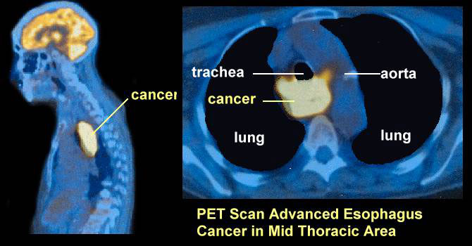
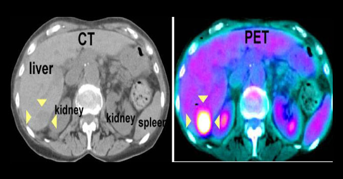
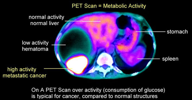
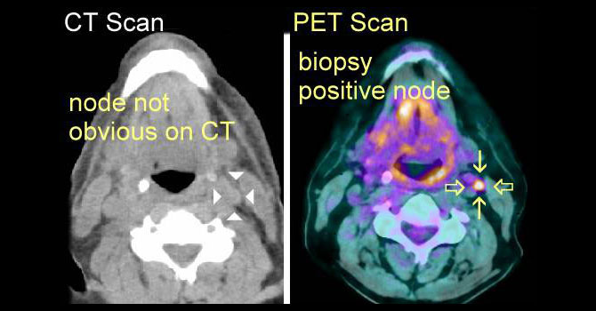
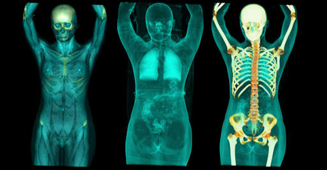
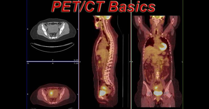
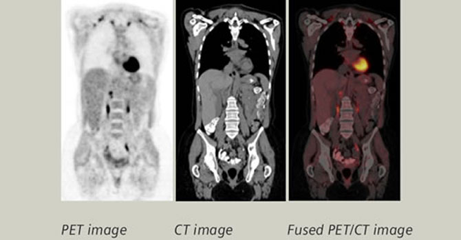
PET CT is a diagnostic imaging test primarily used for detection of cancer (whole body) and several other ailments. The PET-CT Scan is a test that is done on a special PET-CT Machine which performs the 2 scans together:
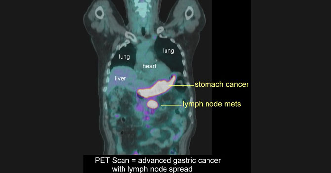
PET Scan: The Positron Emission Tomography or PET Scan component marks most actively upataking cells in the body. During the test, a small dosage of a Radioactive Tracers is injected into the patient. This molecule is an isotope which is actively uptaken by most active cancer cells in the body. The uptake of the tracer is measured by the PET scan component of PET/CT scan, to highlight and mark most cancers.
CT Scan: The Computed Tomography or CT Scan is used for anatomical localisation of the body. A diagnostic quality CT Scan is performed along with the PET scan for the anatomical evaluation of the tests. Prior to the scan, all patients are given oral contrast (positive/negative) to drink. During the scan, intravenous contrast is injected (unless creatinine is high / history of prior allergy / referring physician’s discretion).
BOOK PET CT
Kindly Note, there is an advance amount deposit for booking your medicine dose and appointment for the PET CT Scan. The earliest available appointment for the scan should be confirmed over the phone or in person with the Centre’s Reception. The contact details are as follows:
CP 2/3, Vishwas Khand -2, Near Fly Over
Gomti Nagar, Lucknow-226010
0522-2308987,88
+91 9458704154 ; 9458701800
srmsfimc@srms.ac.in
Preparation for Test Day
Information about Scan Procedures
Information after the PET-CT scan
- You can go home and get back to your daily routine. You can take your food and all your medicines.
- Adequate amount of fluid / water has to be consumed and urine has to be passed out so that the remaining medicines in the body come out.
- If patients are admitted in a hospital, they can be admitted back to their ward.
Important information for PET-CT Scan
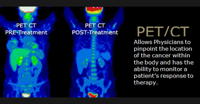
One stop investigation that helps in staging, re-staging, assessing therapy response and helps in customizing management in patients suffering from various types of cancer. Also useful in cases of PUO, latent infection/inflammation etc. scan may be done with contrast/without contrast.
Requirement: fasting blood glucose level less than or equal to 160-165 mg/dl. Serum Creatinine reports
Preparation:
Fasting not less than 6 hrs.
P.S: For diabetics, meet doctor on the day of appointment regarding instructions for management of blood glucose. They must inform department staff about their status on arriving on the day of investigation.
Book now
One stop investigation that helps in staging, re-staging, assessing therapy response and helps in customizing management in patients suffering from carcinoma Prostate or biochemical recurrence in post-op patients. Scan may be done with contrast/without contrast
Requirement:No strict Blood glucose control required.
Preparation:
NPO 4hrs
Book now
Scan done to assess viability of heart muscles (LV myocardium). This is followed by rest MIBI cardiac scan to which FDG PET-Ct cardiac images are compared to.
Requirement:fasting blood glucose level less than or equal to 160-165 mg/dl.
Preparation:
Fasting not less than 6 hrs.
P.S: For diabetics, meet doctor on the day of appointment regarding instructions for management of blood glucose. They must inform department staff about their status on arriving on the day of investigation.
Book nowUsing Flow Motion Technology to
Overcome Today’s PET/CT Challenges

Until now, PET examinations have been performed in sequential bed positions, alternating between acquisition and patient table motion. As such, image quality and quantitative accuracy have always been restricted, and scans often resulted in higher dose and greater patient anxiety. Powered by Siemens’ revolutionary FlowMotion technology, Biograph mCT Flow is the world’s first PET/CT system to eliminate the demand for stop-and-go imaging.

With Flow Motion examinations, parameters such as speed, image resolution and motion management can be easily adjusted to the precise dimensions of organs and routinely

FlowMotion technology eliminates overlapping bed acquisitions while maintaining uniform noise sensitivity across the entire scan range, enabling

FlowMotion allows easy anatomy-based scan planning where only the desired area is scanned.

Biograph mCT Flow is the first-and-only PET
continuous sense of scan progress, offering the
Unique Features
Finest Volumetric Resolution
Biographm mCT Flow with its OptisoHD detection system optimizes each component of the imaging chain.In addition to that, this comes with single source dual-energy CT capabilities for improved lesion contrast.
Virtually Freeze Motion
Through an innovative combination of hardware and software, HDoChest virtually freezes respiratory motion, enabling full HD lesion detection and accurate standard uptake value (SUV) quantification.The five-year survival rate for NSCLC may more than double if caught earlier, benefiting up to 85% of lung cancer patients. 3FlowMotion simplifies integration of HDoChest into every scan.
Precise Organ Imaging
Examination parameters such as speed, image resolution and motion management can be easily adjusted to the precise dimensions of organs.
Hi-Rez Increases Accuracy
Improved accuracy can change patient management in up to 40% of head and neck cancer cases.4 FlowMotion technology additionally provides simple incorporation of Hi-Rez into the PET scan protocols. This PET reconstruction with the industry’s highest1400x400 matrix can increase resolution potentially resulting in improved detection.
Oncological Applications
Non-Oncological Applications
Neurology
Cardiology
Assessment of myocardial viability in patients with Ischaemic Heart Disease and poor left ventricular function being considered for revascularization in combination with perfusion imaging with sestamibi/tetrofosmin/ thallium.
Infection imaging
Sarcoidosis
Assessment of activity and distribution of disease at baseline in highly selected cases where there is diagnostic uncertainty using conventional imaging (e.g. suspected cardiac sarcoidosis). Assessment of disease response where other measures to monitor response are unhelpful and/or in patients with disease resistant to treatment.
Vasculitis
Evaluation of suspected vasculitis in selected cases; for example, to determine the extent & distribution of the disease activity.
Pyrexia of unknown origin (PUO)
To identify the cause of a PUO where conventional investigations have not revealed a source.


