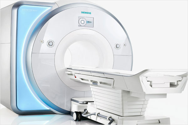
MAGNETOM Skyra
3.0 TESLA MRI with 48 channels
The first Silent MRI System of Uttar Pradesh
MAGNETOM Skyra -The 48 channels,72 cm wide bore 3 TESLA MRI, MAGNETOM Skyra maximizes the potential of 3T, taking MRI to the next level. This scanner delivers impeccable image quality
MRI uses a strong magnetic field and radio waves to create detailed images of the organs and tissues within the body. An MRI scan can be used to examine almost any part of the body, including the brain, spinal cord, bones and joints, breasts, heart and blood vessels, internal organs, such as the liver, womb or prostate gland.
Advantages
The MRI scan procedure is safe and radiation-free, implying that it is safe even for pediatric and repetitive studies. They’re frequently used to diagnose issues with your joints, brain, wrists, ankles, breasts, heart, blood vessels. The advantages of MR Imaging scan are:
- MRI scanning is painless and does not involve x-ray radiation.
- It provides valuable information on glands and organs within the abdomen, and accurate information about the structure of the joints, soft tissues, and bones of the body.
- There is no involvement of any kind of radiations in the MRI, so it is safe for the people who can be vulnerable to the effects of radiations such as pregnant women or babies.
- MRI Scan can detect deformities that might be unclear by bone with other imaging methods.
- One big advantage to an MRI scan is that there is no exposure to radiation as in the case with X-rays, CT scans, and PET scans.
Preparation & Procedure
On arrival at the hospital, doctors may ask the patient to change into a gown. As magnets are used, it is critical that no metal objects are present in the scanner. The doctor will ask the patient to remove any metal jewellery or accessories.Once the patient has entered the scanning room, the doctor will help them onto the scanner table to lie down.The table then slides into the cylinder. An intercom inside the MRI scanner allows you to talk with the radiography staff. It is important to lie very still. Movement will blur or distort the pictures. Women should always tell their doctor and technologist if they are pregnant or planning to get pregnant. There might be a risk of harm for the fetus during an MRI scan. When the MRI scan is complete, you may be asked to wait while the radiologist checks the images in case more are needed. The entire MRI procedure may take 30-60 minutes.
Time Taken For MRI Scan Reports
- The Timing of the Scan: Depending on when you have your MRI stand, it may take longer for you to get your results.
- If it’s an emergency: You’ll usually get MRI results more quickly if you underwent the MRI for an emergency.
- The Size of the MRI Scan: Another factor that will play a role is the size of the MRI scan. For example, if you are getting an MRI of your entire body, it is going to take the radiologist longer to read this scan. On the other hand, if you are only getting an MRI scan of your head, that it might be read more quickly.
MRI Reports will be available within 24-48 hours of the procedure in the maximum cases.
Book now
SRMS FIMC System delivers exceptional quality and speed in 3T MRI
MAGNETOM Skyra with the groundbreaking integration of Tim® 4G and DotGo® sets a new standard of efficiency, ease of use, and care which will helps harness a new level of patient comfort.
- TIM 4G – High coil density enables high SNR, resulting in excellent image quality and higher PAT factors for faster exams.
- DotGo – The unique software ensures higher consistency.
MAGNETOM Skyra at a glance
Services Maximize 3T. Every case. Every day
> 1 minute
Exam-time variation
Up to 40%
Faster MRI exams*
up to 97%
Reduction in sound pressure
70cm
Open bore
The first Quiet MRI System
with both NEURO and ORTHO applications
Siemens-unique Caipirinha : Ensures faster exams that play a major role when patients have to hold their breath.- With CAIPIRINHA we reduce breath hold times from 20s and more to 12-14seconds without compromising image quality.
HEAD / NECK
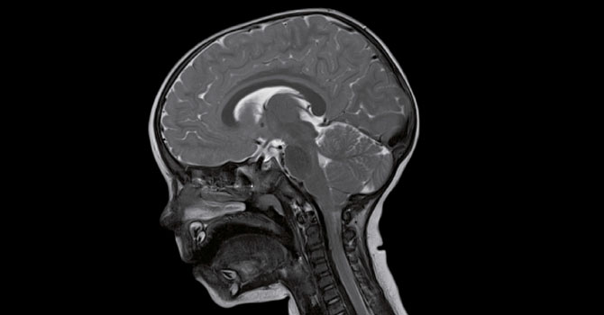
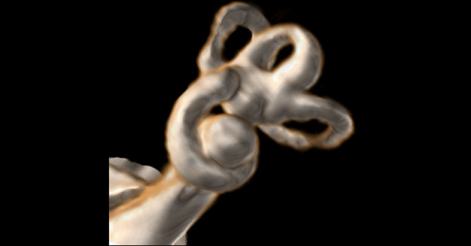
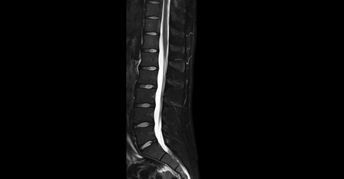
With excellent SNR, the Head/Neck 64 coil reveals previously hard-to-recognize details in the brain, inner ear, orbits, skull base, neck and spinal cord – improving insights in neuroanatomy while improving scan time and resolution.
NEUROLOGY
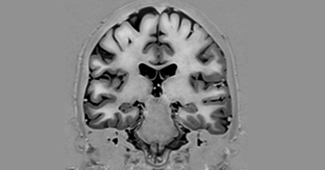
Tim 4G delivers benefits in neuroscience in both research and clinical routine – thanks to increased SNR and a new MR architecture
PEDIATRICS
Ultra-high density, light-weight Tim 4G coils enable faster examinations, helping reduce the need for sedation and rescans in pediatrics.
ORTHOPEDICS
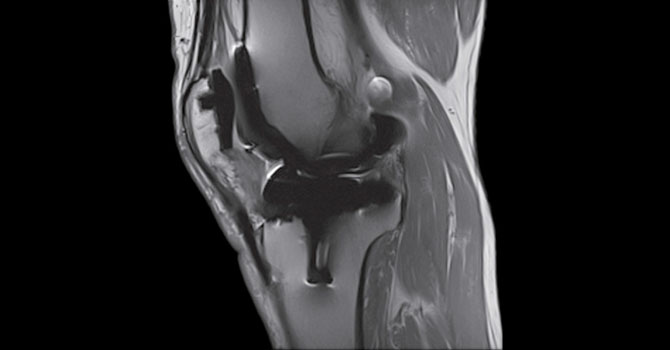
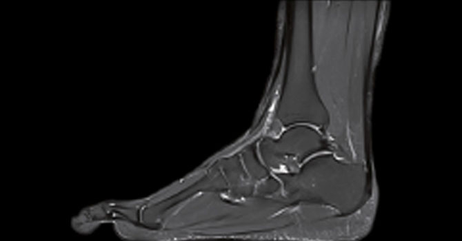
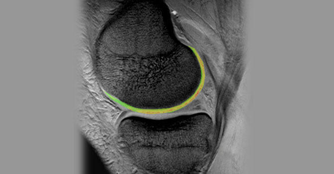
Tim 4G’s ultra high-density coils for MSK imaging maximize SNR and anatomic coverage.
ANGIOGRAPHY
Tim 4G’s new ultra high-density coils and 205 cm scan range allow you to perform high-resolution whole-body angiography easily and without repositioning the patient.
CAIPIRINHA
Address patients with limited breath-hold capacity with Siemens’ unique CAIPIRINHA application and ultrashort breath holds
Liver Lab
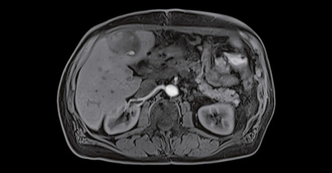
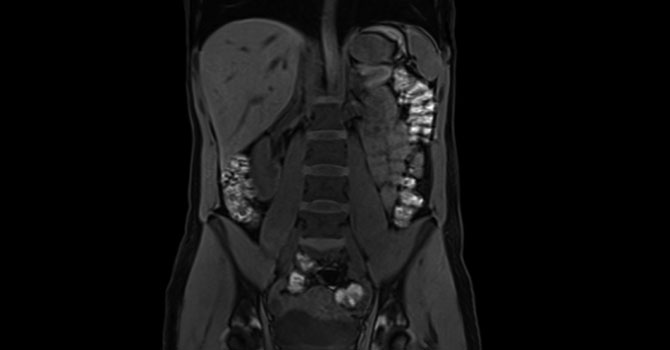
For absolute quantification of liver which is fully automated with a scan time of maximum 5 minutes. Moreover this enables a total non invasive measurement in screening the Liver.
FREEZE it
Consists of TWIST-VIBE and StarVIBE.
TWIST-VIBE
is an Ultrafast, dynamic liver imaging for always the right contrast timing, leading to additional findings without missing any arterial phase. This avoids the repeat scanning thereby increases the patient comfort and reduces the contrast administration.
StarVIBE
is a Free-breathing contrast-enhanced liver imaging to open up body imaging for a broader patient population. This is absolutely unique and comes very handy for patients like Paediatric, Adults who are incapable of holding the breaths (Asthmatic patients), Very sick patients and the Old population. With FreezeITyou we achieve a Breathing artifacts free imaging all the time.
BODY
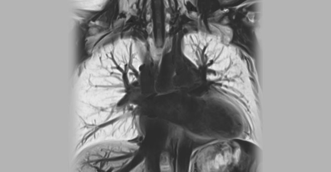
Tim 4G offers high-channel body imaging due to the combination of the ultra-high density body and spine coils.
PROSTATE MRI
With the high-density spine and body coils alone , Tim 4G delivers excellent flexibility in multiparametric imaging of the prostate in terms of morphology, physiology and function.
MR Elastography
provides the ability to noninvasively assess relative stiffness of liver tissue to improve diagnosis for treatment decisions, especially in liver fibrosis. We can now scan the donor within 6 minutes and come to a clinical decision.
WARP
Serve the growing population of patients with MR-conditional metal implants. Advanced WARP: Serve the rapidly-growing patient populations that have artificial joints. The benefits are substantial: infections can be diagnosed earlier and there’s a significant gain in image quality for any MR indication.
