CT Scan

Fastest & Healthiest

CT Scanner in Lucknow

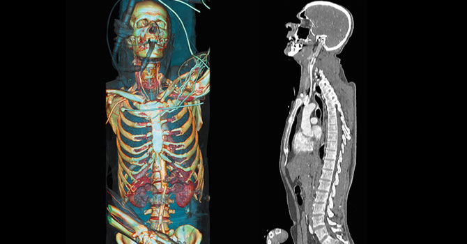
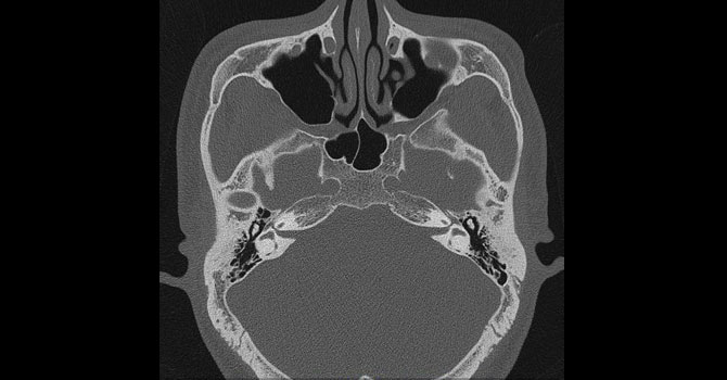
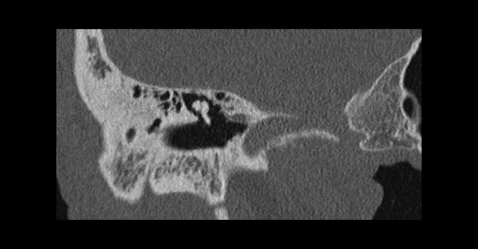
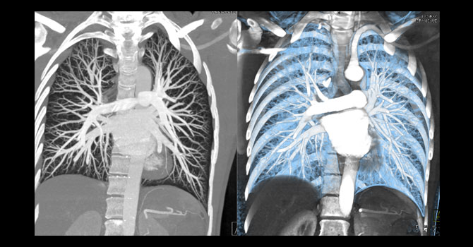
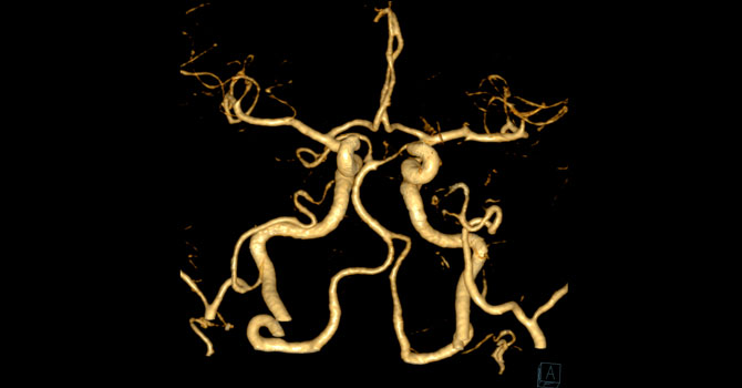
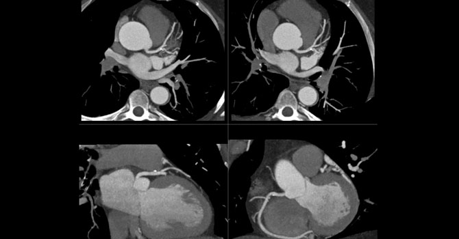
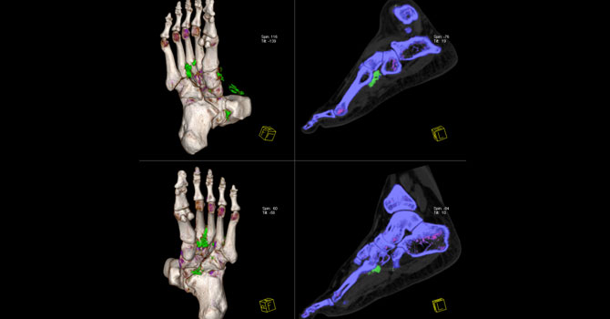
A CT scan or computed tomography scan (formerly known as computed axial tomography or CAT scan) is a medical imaging technique used in radiology to get detailed images of the body noninvasively for diagnostic purposes. The personnel that perform CT scans are called radiographers or radiology technologists. A CT scan uses X-rays and computers to produce images of a cross-section of your body. It takes pictures that show very thin “slices” of your bones, muscles, organs and blood vessels so that healthcare providers can see your body in great detail.

Advantages

  • MRI scanning is painless and does not involve x-ray radiation.
  • most accurate tools for examining the chest, abdomen and pelvis because it provides detailed, cross-sectional views of all types of tissue
  • Determining when surgeries are necessary
  • CT examinations are fast and simple; in emergency cases, they can reveal internal injuries and bleeding quickly enough to help save lives.
  • It rapidly acquire images and provide clear and specific information.

Preparation & Procedure

You may need to wear a gown during the scan. Metal objects including jewelry, eyeglasses, dentures and hairpins may affect the CT images and should be left at home or removed prior to your exam. You may also be asked to remove hearing aids and removable dental work. you should not eat or drink anything for several hours before your scan, especially if a contrast material will be used in your exam. You should inform your physician of all medications you are taking and if you have any allergies. Also, Inform your doctor of any recent illnesses or other medical conditions and whether you have a history of heart disease, asthma, diabetes, kidney disease or thyroid problems.

During the test, you will lie on your back on a table (like a bed).The bed slowly moves into the doughnut-shaped scanner. At this point, you will need to stay as still as possible because movement can create blurry images. The scanner takes pictures of the area the healthcare provider needs to see. Unlike an MRI scan, a CT scan is silent. When the exam is over, the table moves back out of the scanner.

Time Taken For CT Scan

You can have a CT scan done in a hospital or an outpatient facility. CT scans are painless and, with newer machines, take only a few minutes. The whole process typically takes about 30 minutes

Book now


The SOMATOM Definition AS Platform

  • World’s first Adaptive Scanner suitable for all body types
  • Fastest CT scanner in Lucknow – 0.33 sec rotation
  • The first system to come with the FAST CARE Platform
  • First scanner to come with automatic kV selection
  • Suitable for scanning children at the lowest dose
  • Dual Energy ready CT
  • The most trusted high-end scanner in the world:
  • 64 slice acquisition / 192 slice reconstruction CT scanner
  • Best image quality with highest resolution
  • Wide Bore (78 cm gantry bore) scanner for comfortable scanning

Industry best 30 Ip/cm ensures the highest image
quality at the lowest dose

QUALITYHIGH END CT WITH THE ABILITY TO PERFORM ALL TYPES OF SCANS
  • All routine CT Applications – CT Head, Abdomen, Emergency, Thorax, Peripheral, Extremities
  • High End Applications – Complete range of Cardiac, Neuro, Oncology and Dual Energy applications


  • Fast 0.33 sec rotation ensures the highest cardiac image quality, Also, Adaptive Cardio Sequence and Spiral ensure that the scanner can overcome any missing heart (ectopic) beats
  • Complete cardiac-vascular package (inclucing Cardiac Functional) with 64 slice acquistion provides all the tools required for reporting cases, including complex cases such as by-pass (CABG) follow-ups and case where the heart doesn’t pump blood well (i.e. with lower LV function)


  • Neuro DSA provides cathlab like Neuro Angios for faster and more accurate
    diagnosis. This allows for easy viewing of eneurysms (that cause brain bleeds and can
    be the cause of sudden death) and stenosis (blockage of the vessels) in the brain.
  • Neuro Perfussion ensures that all storke cases can be handled in the golden hour
  • Sharp imaging and high detail in smaller structures like Inner Ear.


  • Perfect synchronization with the PET allowing for multi-timepoint comparison of both PET and CT images before and after scanning. Also included are RECIST and WHO measurements to report according to the global standard
  • 0.33 mm resolution allows accurate display of even very small vessels
  • Entire thorax abdomen can be examined without artifacts-e.g. to check on liver lesions
  • CT guided interventions for biopsy of tissue (to check of its cancerous or not)


syngo.via: Multimodality solution with CT, MR and PET applications

  • Pre-processing: All cases are pre-processed within seconds-so that the cases are ready to report for scanning. E.g. in Cardiac cases, a double click on the case opens up multiple views, including cardiac VRT with blood pool removal, all the three planes and VRT with all major vessels labelled, Curved MPR’s of all major vessels etc.
  • Complete patient history: With the same patient name/ nomenclature, the user can look across multiple scans conducted across the diffrent modalities for
  • Image Fusion
  • Multi timepoint comparison
  • Follow up cases
  • Check on Efficacy of interventins/surgeries
  • Applicatins such as Spine and Rib lebelling, Bone removal etc support the clinician in quick and accurate reporting


Wide Bore (78cm) CT provide the flexibility to handle & also provides much higher patient comfort

  • Very heavy patients
  • Emergency cases
  • Cancer treatment (Radiaton Therepy) planning
  • CT Guided Interventions

FAST (Fully Assisting Scanner Technologies), including FAST Planning and FAST Spine provide
great workflow advantages at the scanner console. These are used to improve accuracy and
reproducibility of scanning and reporting.

CARD Applications such as CARD Dose 4D and CARD kV reduce dose for the patient while
providing the optimum image quality


Image Gallery

  • MRI
  • MRI
  • MRI
  • MRI
  • MRI
  • MRI
  • MRI
  • MRI
  • MRI
  • MRI